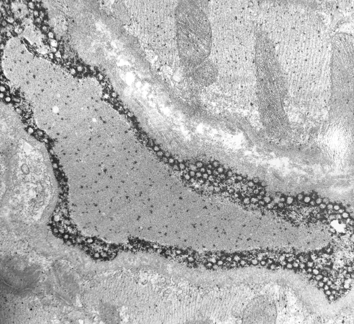
Capillary in cardiac muscle reacted for Purine Nucleoside Phosphorylase
Resin embedding for light microscopy provides greatly improved cellular definition compared to paraffin embedding, and for this reason is now widely used in diagnoses; particularly of Renal disease, Lymphomas and bone marrow trephines, as well as research.
Many acrylic resins are not suitable for EM and the epoxy resins used for EM are not easily stained for light microscopy. LR White however can be used for both purposes and a lymph node for example (12x10x3mm) can be processed, cut and stained for light microscopy. The same block can then be trimmed, cut and stained for electron microscopy.
LR White can also be used for the histochemical demonstration of some of the more resistant enzymes, and for the immunocytochemical demonstration of intracellular immunoglobulins.
For those laboratories already using an acrylic resin e.g. HEMA or Glycol Methacrylate no alteration need be made to the current processing schedule, but we have laid out here a 'typical' schedule for LR White as guidance for its use.
Fixation:
No change from normal fixation need be made if LM analysis only is required from the final blocks (Neutral Buffered Formalin recommended). If, however EM is required subsequent to LM then we have found the use of aqueous formalin in a phosphate buffer pH 7.2 with 2.5% w/v sucrose is the best compromise. Glutaraldehyde-formaldehyde mixtures may lead to very pale staining with haematoxylin and patchy eosin, whereas normal formalin fixation gives unacceptable EM structure. For the dual LM/EM application, osmium tetroxide should be avoided due to its effect on many LM stains but 1% phosphotungstic acid (w/v) in the first absolute ethanol step of dehydration improves electron contrast without adversely affecting most LM stains. If this does not provide adequate electron density, then 'staining' of ultrathin sections can be carried out with osmium (a brief exposure to 1% aqueous osmium tetroxide or osmium tetroxide vapour on a copper grid) or lead citrate.
Dehydration:
A graded ethanol series is the method of choice when using LR White. Acetone acts as a radical scavenger in the resin system and traces of acetone left in the tissue at curing can interfere with polymerisation.
Infiltration:
The extremely low viscosity of LR White allows the use of short infiltration times, but these will depend on the size of the tissue. Infiltrated tissue will become translucent and sink to the bottom of the container.
A typical dehydration and infiltration schedule for a block (12x10x3mm) on a mixer would be:
| 1. Two changes 70% alc | 30 mins each |
| 2. Two changes Abs alc | 30 mins each |
| 3. Infiiltrate with LR White at room temperature 2-3 changes | 60 mins each or leave overnight |
Polymerisation:
Either heat or cold curing can be used for LM, with the latter giving slightly better cutting and staining qualities. When cold curing it is important to cool the moulds in a bath of cold water, during polymerisation, to disperse the heat produced by the exothermic reaction, but it is not necessary to exclude oxygen from the surface of the curing block.
Some polymerisation problems have been experienced when embedding very flat pieces of tissue which stick to the base of the embedding mould. The way to avoid this is to smear the base of the mould with accelerator before adding mixed resin and allow the tissue to sink to the base of the mould rather than applying pressure.
When thermal curing it is important to limit the contact of oxygen with the resin while polymerisation occurs. The most convenient way of achieving this is to use gelatine capsules (available from the Agar Scientific) for small pieces of tissue. Fill up to the brim and slide the other half of the capsule on. For larger specimens the surface of the resin must be covered, and one convenient method is to utilise the JB-4 type moulds, one being used as a lid for another, or to polymerise in a nitrogen environment. Polymerisation time and temperature are fundamental to the physical characteristics of the final block, and to a much greater extent than with under-cured epoxy systems.
We strongly recommend a temperature of 60°C ± 2°C for a period of 20-24 hours. Some ovens are not capable of controlling temperature so closely and if faced with over brittle blocks this is the first parameter to check.
Resin may be used straight from the refrigerator and has a very low toxicity in both monomeric and polymerised states unlike epoxies (see Proc. Roy. Mic. Soc. (1981), 16, Pt.4, p. 265-271). The cold cure accelerator does have some toxic risk and contact with skin and eyes should be avoided.
For cold curing the accelerator should be used at one drop per 10ml of resin and this should case polymerisation in 10-20 minutes. If polymerisation occurs faster than this, we recommend either more careful metering of the one drop of accelerator or a higher volume of resin per drop of accelerator.
Cutting and mounting:
Although it is possible to cut LR White on a standard microtome with a steel knife the method of choice would be to use a heavy-duty motorised microtome, and glass (Ralph type) knife.
LR White can cut as thin as 0.25μm on some microtomes, but it is very difficult to obtain a satisfactory stain intensity with anything other than toluidine blue at this thickness, simply because there is so little tissue present in the sections.
For haematoxylin and eosin staining as well as most other routine stains we recommend sections of 2- 3μm. It is of course possible to cut thicker (up to 15 or 20μm) if required.
Blocks can be cut dry, the sections picked up and floated out on 30-40% acetone on a hot plate @ 60-70°C, and then allowed to dry at this temperature. For hard tissues, and blocks which contain a combination of hard and soft tissues, such as marrow trephines, the above mentioned floating out fluid is recommended, again on a hot plate @ 60-70°C.
To 20ml acetone add 0.5ml benzyl alcohol mix then make up to 50ml with distilled water. A section adhesive such as egg albumin, can be added to this if required.
Section staining:
Most routine stains give good results on tissue embedded in LR White resin using standard times and temperatures although it may occasionally be necessary to extend some staining times e.g. Methyl Green Pyronin. Stains made up in ethanol or methanol should be avoided as these solvents soften the resin and may remove sections from the slide. Dehydration of sections through graded alcohols after staining should also be avoided. Sections should be blotted, air dried and then mounted in a resinous mounting medium.


