Gold nanoparticles are utilized widely in life science applications due to their biocompatibility and ease with which they can be conjugated with organic molecules. Control over nanoparticle size and structure is critical to providing reliable and reproducible results. Gold nanoparticles of different sizes in solution are readily available, however these nanoparticles are often contaminated with hydrocarbons as a result of the chemical synthesis and clump together in solution.
The NL50 offers an alternative route to produce monodisperse and ultra-pure gold nanoparticles which are free of contaminants. The NL50 generates the nanoparticles from solid source material with 99.999% purity. The nanoparticles are generated in vacuum using a process called magnetron sputtering where atoms are knocked from the source material by energized gas ions. These atoms clump together to form nanoparticles. The size of the nanoparticles can be controlled by changing the process parameters of gas flow and magnetron power inside the NL50. The NL50 can produce nanoparticles from any conductive material, including gold, silver, titanium, copper, platinum and nickel. The following video explainer shows how the NL50 works.
The ultra-pure nanoparticles are extracted from the nanoparticle generation zone of the NL50 into the sample chamber where they land on the substrate. The NL50 can deposit nanoparticles onto any solid substrate including glass, plastic, metal, which has been loaded by the operator through the transparent sample door.
Gold nanoparticles were deposited on holey carbon grids (supplied by Agar Scientific) for TEM analysis. The TEM analysis was carried out by Max Astle and Rhys Lodge at the Nottingham Nanoscale and Microscale Research Centre (nmRC) using a JEOL 2100F FEG-TEM microscope operated at 200 keV.
Figure 1 shows TEM images of the gold nanoparticles, confirming that the nanoparticles are discrete, crystalline and form the well-recognised icosahedral structure. Figure 2 shows that the nanoparticles have tight size distribution centred around 22nm. In the high-resolution image, shown in figure 3, the individual gold atoms are clearly visible.

Figure 1: Gold Nanoparticles prepared in vacuum in the NL50
Figure 2: Size distribution of gold nanoparticles from Figure 1
Figure 3: High-resolution image of gold nanoparticle showing icosahedral structure
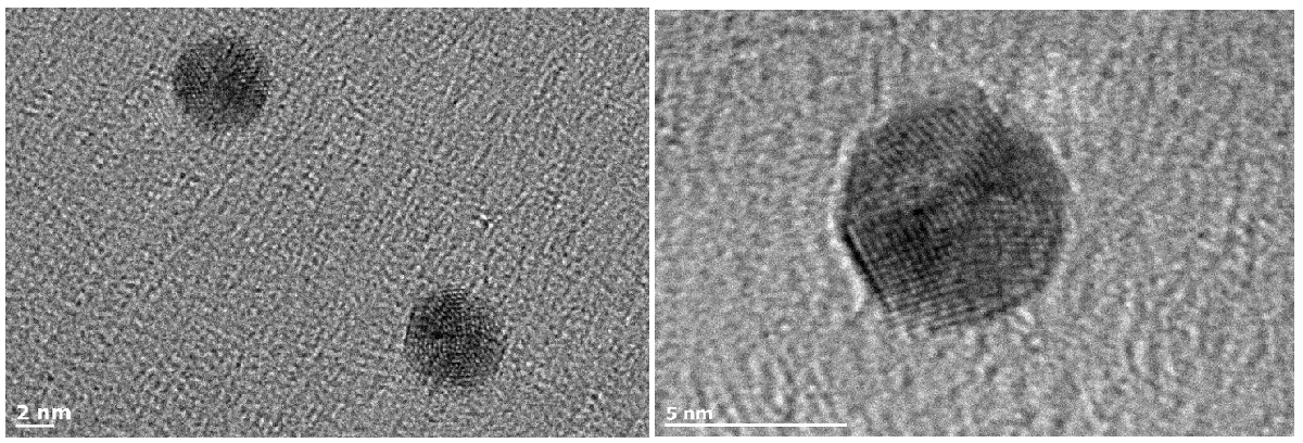
Smaller nanoparticles can also be produced in the NL50 by changing the process conditions. Figure 4 shows TEM images of smaller nanoparticles with sizes of 4.5 and 6.5nm. The smaller nanoparticles are also crystalline but the shape is more spherical than the larger nanoparticles.

Figure 4: Small gold nanoparticles generated in the NL50 with diameters of 4.5nm (left) and 6.5nm (right)


